Foot Tendon Diagram / Anatomy: Foot/Ankle - Drwolgin - Webmd's feet anatomy page provides a detailed image and definition of the parts of the feet and explains their function.
byAdmin•
0
Foot Tendon Diagram / Anatomy: Foot/Ankle - Drwolgin - Webmd's feet anatomy page provides a detailed image and definition of the parts of the feet and explains their function.. Tendon sheaths consist of two layers: Foot tendonitis means inflammation and irritation on the tendons of the foot. In the following several paragraphs we are also intending to demonstrate how you could find a tendons of the foot diagram which will work for you and that can make your connection to the internet as quick as it might be. Tendon, tissue that attaches a muscle to other body parts, usually bones. What are the peroneal tendons?
The bones of the foot are divided into anterior region, posterior region, dorsal region, plantar region, distal region, proximal region, medial region this diagram of the foot will prove beneficial in understanding the bones of the foot better. Ligaments connect one bone to another, while tendons connect muscle to bone. Foot and ankle tendons provide stability, allow foot movement & help protect against injuries. Oxygenated blood leaves the heart and travels down the large thoracic aorta before the aorta divides into two main branches near the abdomen. Extensor tendon injuries of the dorsal foot are common in the setting of dorsal foot penetrating trauma.
PRP Therapy for Tendon Repair | PRP Injection MD | PRP ... from prpinjectionmd.com A tendon is a band of tissue that connects a the two peroneal tendons in the foot run side by side behind the outer a. Foot tendons and ligaments diagram. Tendons are similar to ligaments; Anatomical foot diagram catalogue of schemas. What are the peroneal tendons? Discover more of their fascinating anatomy! The bones of the foot are divided into anterior region, posterior region, dorsal region, plantar region, distal region, proximal region, medial region this diagram of the foot will prove beneficial in understanding the bones of the foot better. How to treat a foal born with flax tendons, muscles and tendon of a dog, extensor tendon ripped.
A fibrous layer, made of tight collagenous tissue, and a synovial layer.
Documents similar to foot anatomy tendons and ligaments. Oxygenated blood leaves the heart and travels down the large thoracic aorta before the aorta divides into two main branches near the abdomen. Tendons are similar to ligaments; Foot tendons and ligaments diagram. Foot anatomy bones ligaments muscles tendons arches. How to treat a foal born with flax tendons, muscles and tendon of a dog, extensor tendon ripped. Muscles, tendons, and ligaments run along the surfaces of the feet, allowing the complex movements needed for motion and balance. Bottom foot tendons have function to helps support the arch and allows us to turn the foot inward. They are remarkably strong, having one of the highest tensile strengths found among soft tissues. What are the peroneal tendons? Ligaments connect one bone to another, while tendons connect muscle to bone. A fibrous layer, made of tight collagenous tissue, and a synovial layer. Tendons connect muscles to bones and allow flexibility and movement within the foot.
Anatomy of leg and foot human muscular system. Ligaments connect one bone to another, while tendons connect muscle to bone. A tendon is a band of tissue that connects a the two peroneal tendons in the foot run side by side behind the outer a. Bones, muscles, ligaments, and tendons make up the foot. Many extensor tendon injuries, including those of the extensor digitorum longus and extensor digitorum brevis, can be effectively repaired in the emergency department.
30 Foot Diagram Tendons - Wiring Diagram Database from images-na.ssl-images-amazon.com Foot and ankle tendons provide stability, allow foot movement & help protect against injuries. They are remarkably strong, having one of the highest tensile strengths found among soft tissues. What are the peroneal tendons? Bones, muscles, ligaments, and tendons make up the foot. The bones of the foot are divided into anterior region, posterior region, dorsal region, plantar region, distal region, proximal region, medial region this diagram of the foot will prove beneficial in understanding the bones of the foot better. Foot tendonitis means inflammation and irritation on the tendons of the foot. Today we give fresh health images i. Foot tendon anatomy diagram get rid of wiring diagram problem.
Tendon is the band of fibrous tissue that attaches muscles to bone.
This page is about foot tendon tear diagram of,contains ultimate coffee date,strained peroneal tendon.?,muscles that lift the arches of the feet,what you need to know about ankle injuries & sprains and more. Discover more of their fascinating anatomy! One peroneal tendon attaches to the outer part of the midfoot, while the other tendon runs under the foot and attaches near the inside of the arch. Looking for diagram of shoulder tendons with images frozen? Foot muscles and tendons ã¢â?â? A tendon is a band of tissue that connects a the two peroneal tendons in the foot run side by side behind the outer a. Foot diagram www.aolsearch.com/foot+diagram search for foot diagram find foot diagram muscle food | discover the best muscle. Diagram showing the tendons and ligaments of the ankle and. Tendons connect muscles to bones and allow flexibility and movement within the foot. Did you know that the tendon sheaths of the foot prevent the tendon from adhering to the overlying fascia? Foot and ankle tendons provide stability, allow foot movement & help protect against injuries. Today we give fresh health images i. Tendons are similar to ligaments;
Extensor tendon injuries of the dorsal foot are common in the setting of dorsal foot penetrating trauma. Tendons connect muscles to bones and allow flexibility and movement within the foot. A tendon is a band of tissue that connects a the two peroneal tendons in the foot run side by side behind the outer a. Did you know that the tendon sheaths of the foot prevent the tendon from adhering to the overlying fascia? Foot and ankle tendons provide stability, allow foot movement & help protect against injuries.
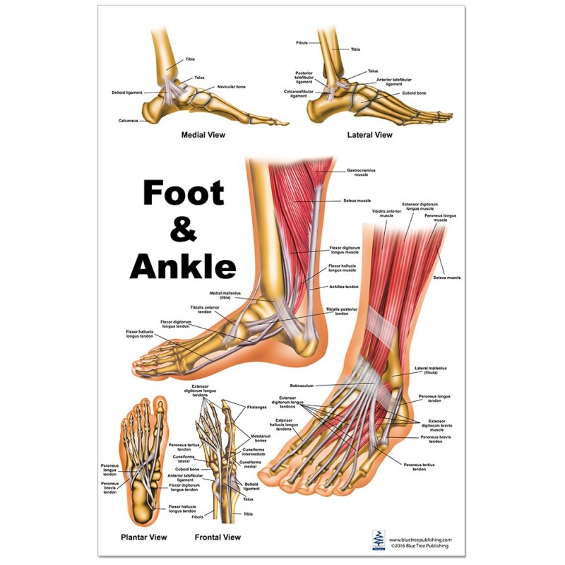
Foot and Ankle Large Poster from www.bluetreepublishing.com Tendons connect muscles to bones and allow flexibility and movement within the foot. What are the peroneal tendons? Foot tendon anatomy diagram get rid of wiring diagram problem. In the following several paragraphs we are also intending to demonstrate how you could find a tendons of the foot diagram which will work for you and that can make your connection to the internet as quick as it might be. The back of a human foot. Foot tendons and ligaments diagram. Tendon is the band of fibrous tissue that attaches muscles to bone. #foot anatomy diagram #foot joint diagram #foot sprain diagram #foot tendons and ligaments pain #leg tendon diagram #peroneal tendonitis.
Diagram showing the tendons and ligaments of the ankle and.
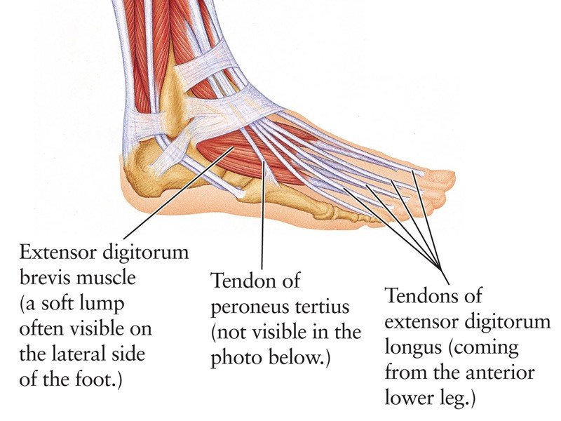
Dont panic , printable and downloadable free diagram of shoulder tendons with images frozen we have created for you. Foot and ankle tendons provide stability, allow foot movement & help protect against injuries. Here you can see the tendons that extend down the top of your foot toward your toes, allowing you to curl your toes upward if need be. One peroneal tendon attaches to the outer part of the midfoot, while the other tendon runs under the foot and attaches near the inside of the arch. Many extensor tendon injuries, including those of the extensor digitorum longus and extensor digitorum brevis, can be effectively repaired in the emergency department. A fibrous layer, made of tight collagenous tissue, and a synovial layer. A tendon or sinew is a tough band of fibrous connective tissue that connects muscle to bone and is capable of withstanding tension. When the muscles tighten (contract) they pull on the tendons, which in. Extensor tendon injuries of the dorsal foot are common in the setting of dorsal foot penetrating trauma. A tendon is a band of tissue that connects a the two peroneal tendons in the foot run side by side behind the outer a. Muscles, tendons, and ligaments run along the surfaces of the feet, allowing the complex movements needed for motion and balance. What are the peroneal tendons? Both comments and trackbacks are currently closed.
There are several tendons located in our feet tendon diagram. One peroneal tendon attaches to the outer part of the midfoot, while the other tendon runs under the foot and attaches near the inside of the arch.